
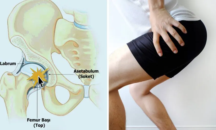
 Hip impingement is caused by increased friction due to a mismatch between the ball-shaped head of the thigh bone (femur) and the socket that holds it (acetabulum). Normally the head of the thigh bone glides easily in all directions within the socket. Certain mismatches in this ball-and-socket mechanism can disrupt smooth movement. Hip impingement syndrome (femoroacetabular impingement, FAI) often shows itself with pain around the hip, a catching sensation and sometimes sharp, stabbing pain (especially when rotating the hip and squatting). Although the exact cause is not fully known, slight developmental irregularities of the joint surfaces may be aggravated by very strenuous activity in athletes, and it is also thought to arise during the development of the hip joint in childhood. FAI is associated with cartilage damage, labral tears, early hip osteoarthritis and low back pain, and is more common in elite athletes and people with an active life. Hip impingement is theoretically one of the factors leading to hip osteoarthritis and one of the causes of early-onset hip osteoarthritis. There are three types of hip impingement!
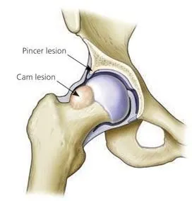 The problem causing impingement can take three forms:
– Pincer type: If the front rim of the acetabulum is longer than it should be, the neck of the thigh bone can strike it when the hip is bent, causing impingement. It is more common in middle-aged women.
– Cam type: If the femoral head, which is normally a smooth sphere, is irregular – i.e. has abnormal bumps – the head jams against the rim of the socket during movements that bend the hip, such as tying shoelaces or pedalling. It is more common in young men.
– Mixed type: There may be a problem in both the pelvis and the thigh bone. In many cases these two forms occur together.
Other conditions can also cause hip impingement. In Legg-Calvé-Perthes disease the femoral head is not adequately supplied because of a circulation problem and the bone tissue dies, causing a deformity. Slipped capital femoral epiphysis is a condition in adolescence characterised by the femoral head slipping off the growth plate. "Coxa vara", characterised by a reduced femoral neck angle due to the growth plates of the thigh bone working at different rates in children, is another cause of hip impingement.
What are the signs of hip impingement?
Particularly when the hip is bent forward and rotated inward (kneeling or crossing the legs), the area at the front-upper part of the acetabulum is pinched. Over time the cartilage and labrum in this area of the hip joint are damaged. Symptoms may start insidiously and increase over time with overuse, with gradually increasing pain at the front of the hip, or patients may present with acute pain due to a labral tear. The pain is felt at the front of the hip and in the groin. Patients may feel pain and catching at the front of the hip and thigh when sitting down and getting up, kneeling or crossing the legs.
How is hip impingement treated?
The first step in treatment is rest and reducing the activities that cause pain. Then exercises to stretch and strengthen the muscles around the hip and physiotherapy methods (muscle strengthening, heat application, etc.) are applied. Painkillers can be used during painful periods. Surgical treatment is needed in patients whose pain persists despite all these measures.
Surgery helps to reduce pain and restore activities in the short term and to halt the process leading to osteoarthritis in the long term. The anatomical differences can be treated arthroscopically or with open methods.
Arthroscopically, the hip joint is examined, the bony bumps causing friction are shaved and, if there is cartilage wear, appropriate procedures are performed. Mini-open techniques have also recently become popular; they allow faster recovery without disturbing the blood supply of the hip. Surgical complications of FAI are rare but include: DVT (deep vein thrombosis – blood clot); infection; hip instability and dislocation; femoral neck fracture; avascular necrosis of the femoral head (bone death due to loss of blood supply); heterotopic ossification (abnormal bone formation in soft tissues); nerve damage (sciatic, LFCN, pudendal, peroneal); non-union (in open surgery); adhesions and scarring.
The information on this page is for general information only and does not replace an examination and diagnosis.