
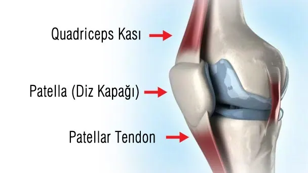
 The kneecap (patella) is a very important part of the knee joint. The knee is the joint between the thigh bone (femur) and the shin bone (tibia), and the kneecap lies at the front of it. The back of the kneecap is covered with smooth, slippery articular cartilage and articulates with a groove in the femur. The quadriceps muscle attaches to the kneecap from above and the patellar tendon from below. When the kneecap slips out of this groove for any reason, this is called a kneecap dislocation.
Why does the kneecap dislocate?
Kneecap dislocations are quite rare injuries, occurring in about 6 per 100,000 people in the general population. The patella may be dislocated from birth (congenital) or the dislocation may develop later. A first dislocation acquired later is called an acute patellar dislocation; repeated dislocations are called recurrent patellar dislocation.
People with patellar dislocation often have underlying developmental bone and soft-tissue problems that predispose to it. Usually because of inborn features, the fit of the kneecap in the joint is poor: the groove the kneecap sits in may be shallow, the ligament that stops the kneecap slipping outward may be loose, or the alignment of the whole leg may be abnormal. In people with these structural factors, dislocation occurs when the body twists inward while the foot is planted on the ground. A first dislocation requires a forceful strain, but in untreated dislocations the soft tissues heal stretched, so recurrent dislocations occur after much simpler movements. Acute patellar dislocation is common under the age of 17 and in girls. In 15% of people with patellar dislocation, someone else in the family has also had one. After a dislocation in one knee, the risk of dislocation in the other knee is quite high.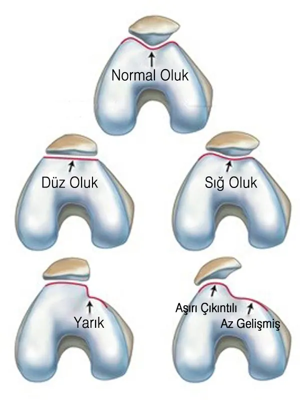 What are the symptoms of acute patellar dislocation?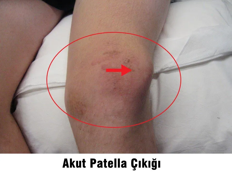
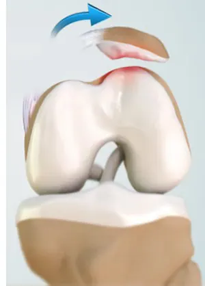 After a sudden twisting movement of the knee, people feel severe pain and feel the kneecap slip to the outside of the knee. Soon afterwards, swelling and limited movement develop due to bleeding into the joint. Often the kneecap goes back into place when the knee is straightened; rarely, patients describe pushing it back themselves.
How is patellar dislocation diagnosed? Does it recur? How is it treated?
Acute patellar dislocation can be diagnosed by examination. If the kneecap is still dislocated, it is put back in place. If the kneecap is in place but tends to slip out, it is assessed with special tests during examination. X-ray, CT and MRI are also used to assess the shape of the kneecap, the depth of the groove it articulates with, its fit in the groove, the condition of the ligaments holding it, and any osteoarthritis.
After a first dislocation, if the kneecap stays in place after reduction and there is no fracture fragment in the joint, the knee is rested to allow the tissues to heal. A rehabilitation programme for muscle strengthening follows. In patients treated without surgery after a single dislocation, the risk of re-dislocation is 17–71%. This risk is higher in people with underlying developmental abnormalities. After a second dislocation, the chance of further dislocations is close to 100%. Kneecap dislocation with an intra-articular fracture, recurrent patellar dislocation and congenital patellar dislocation are treated directly with surgery. Recurrent dislocations can cause irreversible damage to the joint cartilage over time, which may result in osteoarthritis at a young age. The aim of surgery is to prevent recurrent dislocations and progressive cartilage damage in the joint.
Which surgical treatments are used for kneecap (patella) dislocation?
All factors that could cause dislocation must be examined and the operation planned according to the findings. Surgical treatment begins with arthroscopic assessment of the structures inside the joint. In acute dislocations, small loose cartilage-bone fragments are removed and large ones are repaired by fixing them back where they came from. The soft tissues holding the patella in place are repaired directly or, if this is not possible, reconstructed with tendons taken from the back of the knee (MPFL reconstruction). In recurrent dislocations, soft-tissue or bone operations that change the direction of muscle pull are performed, and when needed bone operations to deepen the groove. Congenital patellar dislocations require relocating the whole muscle group at the front of the thigh and, if needed, lengthening the tendon. Sometimes several of these methods are combined.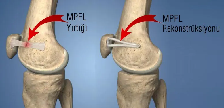 When personalised surgery is performed with modern techniques, the success rate is high. The risk of remaining stiffness or re-dislocation is below 5%. Appropriate treatment prevents cartilage damage from progressing, and the great majority of patients can return to daily life and sport without problems.
The information on this page is for general information only and does not replace an examination and diagnosis.