
Avascular necrosis is a disturbance of the blood supply to the femoral head (the ball of the hip joint) arising from various causes. As a result, over time the bone dies, the femoral head loses its shape, and eventually osteoarthritis (arthrosis) develops in the hip.
It is a disease seen especially in young and middle-aged people that largely requires surgical treatment. Statistics show that it is 8 times more common in men than in women and generally affects people under 50. In our country an estimated more than 3,000 people a year are diagnosed with avascular necrosis.
The disease occurs in the femoral head because only a limited number of vessels supply it. If blood does not reach this area, bone renewal stops, bone is lost, and over time this part of the bone starts to die, breaks up and damages the cartilage over it. As a result, when avascular necrosis develops, the femoral head collapses, loses its roundness and starts to flatten. The load on the hip joint causes this flattening and structural breakdown. The flattening disrupts the fit between the femoral head and the socket; the mismatched surfaces damage each other, leading to osteoarthritis and pain. Total hip replacement, used in the advanced stages, is a very successful operation, but because it is not suited to a highly active lifestyle, wear and loosening may require repeated operations later in life. Preserving the hip joint with interventions in the early stages of the disease is therefore very important. The success of medical and hip-preserving surgical treatments performed before the femoral head collapses ranges from 60% to 80%.
What causes avascular necrosis?
Avascular necrosis has many possible causes, and sometimes it occurs with no identifiable cause. Basically, anything that disturbs the blood circulation of the hip can lead to avascular necrosis; it mostly occurs after a hip fracture or dislocation or fractures of the femoral neck. After such injuries it can take months – sometimes up to 2 years – for avascular necrosis to appear. Long-term steroid treatment, diabetes, sickle cell anaemia, radiotherapy and chemotherapy during cancer treatment, kidney disease, alcoholism, gout, Gaucher disease, clotting disorders, pregnancy, thalassaemia, myeloproliferative diseases, organ transplantation, thrombophilia, smoking, AIDS and decompression sickness (the bends) are also conditions that can cause osteonecrosis.
The reduced blood flow in the femoral head has been explained by several mechanisms: impaired blood flow due to damage to vessels from a fracture or dislocation; fat cells blocking capillaries due to steroid use; or clots forming in capillaries due to a clotting disorder.
What are the symptoms of avascular necrosis?
The most typical symptom is pain. Pain that starts as a mild ache in the groin increases over time. It may be felt in the groin or at the front or back of the hip. Patients limp, walk with difficulty and their walking distance shortens. As the joint deformity increases the pain increases and becomes chronic, and eventually even walking and taking steps become difficult. As the joint cartilage deteriorates, some patients start to hear a "click" during movement, which may be due to a loose fragment in the joint. Over time, as osteoarthritis develops, movements become restricted. Usually the first movement lost is inward rotation of the hip: when the patient pulls the leg towards themselves, the foot turns outwards.
How is the stage of avascular necrosis determined?
Today the most widely used grading systems are based on imaging findings.
(Modified Ficat classification):
| Stage | Criterion |
|---|---|
| I | Plain X-ray normal; abnormal MRI and scintigraphy findings |
| II | Sclerotic and cystic changes present |
| III | Subchondral collapse (crescent sign) and/or step in the cartilage surface |
| IV | Flattening and deformity of the femoral head |
| V | Narrowing of the joint space |
| VI | Signs of osteoarthritis in the joint |
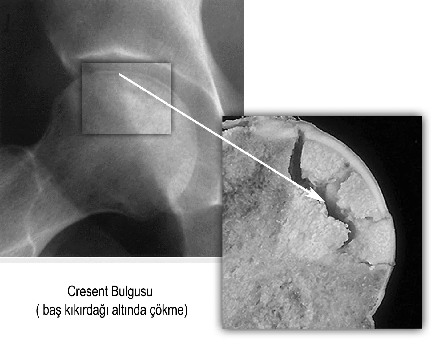
Diagnosis and treatment of avascular necrosis
The most dangerous aspect of avascular necrosis is that it progresses insidiously at first and, because it does not affect quality of life early on, is usually not taken seriously. However, after a short time the symptoms become increasingly severe.
In the early stages of osteonecrosis, X-rays show no signs even though the patient has hip pain. For this reason, hip pain in patients with risk factors for avascular necrosis should be taken seriously, and an MRI must be taken if there is any suspicion. MRI is the best method for early diagnosis; it is very sensitive even when avascular necrosis affects only a small area.
Treatment and assessing the risk of hip osteoarthritis
The first aim of treatment is to make an early diagnosis, identify the cause and remove it. However, by the time the diagnosis is made it is usually too late for this. The second aim in treating avascular necrosis of the femoral head – a difficult problem – is to preserve the existing anatomy (the shape of the hip). Therefore, as well as patient factors such as age, general health and other illnesses, it is important to determine the stage of the disease and the location and size of the affected part of the head. Today we widely use the modified Kerboul classification, which determines the risk according to the amount of necrosis in the head on MRI. The angles of the necrotic area on the mid-sagittal (A) and mid-coronal (B) MRI images are added together:
- less than 190 degrees: low risk
- 190–240 degrees: medium risk
- more than 240 degrees: high-risk group.
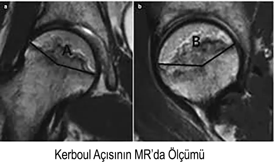 In an article published in 2006 in J Bone Joint Surg Am., based on 5-year follow-up of 37 patients with early-stage osteonecrosis, none of the patients with a total necrosis angle below 190 degrees developed collapse of the head, while all patients above 240 degrees did. Among patients between 190 and 240 degrees, 50% developed collapse.
After determining the stage and risk, the main treatments are:
1- Painkillers and offloading (using a walking stick):
Although in the initial stage offloading and exercises have sometimes been seen to heal the affected part of the bone, they are not a solution in the medium and long term. Painkillers and anti-inflammatory treatment can reduce symptoms but cannot restore the blood supply of the femoral head or reverse avascular necrosis. Unfortunately, therefore, it is not possible to say that this disease responds to medication.
2- Core decompression and/or bone grafting:
This is an operation to increase the bone's blood supply by drilling a tunnel from the femoral neck into the head. There are two techniques: a single 1 cm hole or several 2–3 mm holes. The aim is to reach the diseased area and help new bone form. In 80% of patients operated on in the early stage (Ficat I–II), the bone improves quickly; hip pain disappears and in about 75% of patients the need for joint replacement later is avoided.
In suitable cases bone grafts can also be placed in this tunnel. Bone grafting was first used to provide mechanical support to the drilled area after core decompression, but its popularity declined when it was shown not to be effective on its own in eliminating the risk of collapse.
3- Bone realignment (osteotomy) operations:
In these operations the femoral neck is cut and angled or rotated, so that a healthy area of the head is brought into contact with the socket instead of the damaged part. They may be preferred in the early stages, when less than 30% of the head is involved and collapse is less than 2 mm. However, when most of the head is involved, the necrosis is progressive, and in older patients who may need a prosthesis later, they are no longer a preferred option.
4- Vascularised bone grafting:
Bone with its blood vessel is taken from the leg or pelvis and transplanted into the femoral neck. These are technically difficult and long operations, preferred in stage 3 with collapse of less than 2 mm.
5- Hip replacement:
This is the preferred treatment if the patient is over 60 and/or in the advanced stage, or if hip osteoarthritis has developed. After surgery the hip pain disappears and the patient can move comfortably. The treatment scheme we accept and apply today can be summarised as follows: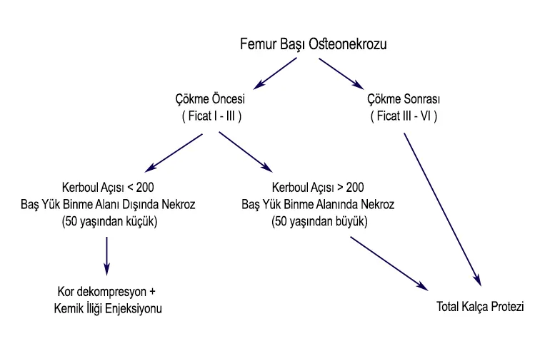
Other treatment methods for avascular necrosis
Because avascular necrosis (AVN) can cause disability, especially in young people, and is seen in patients with active lives, surgery or a hip replacement is not something they want. Surgery means a demanding and difficult period for the patient. For this reason patients try many alternative treatments, as described below. However, there is not yet an accepted, shared scientific opinion on how effective these methods are and at which stage.
Depending on the stage of the disease, improvement in this painful condition may be seen with ozone therapy, hyperbaric oxygen therapy, prolotherapy, acupuncture and herbal treatments as alternatives to surgery.
Hyperbaric oxygen therapy and ozone therapy:
Hyperbaric oxygen therapy (HBOT) involves breathing pure oxygen in a closed pressure chamber. It is used in early-stage avascular necrosis of the femoral head. HBOT alone or combined with surgery has been reported to give successful results in some cases. Its aim is to prevent cell death by delivering more oxygen with less blood to tissue whose blood supply is impaired. HBOT also supports the growth of new vessels and of cells involved in the body's defence, and is used in ischaemic diseases caused by vessel damage or blockage. In ozone therapy, a certain amount of the patient's blood is taken into a bag, enriched with oxygen and returned through a vein. The aim is the same.
Prolotherapy is based on increasing blood flow to the damaged area by injecting proliferative solutions (dextrose) there; it stimulates the body to heal itself by calling repair cells to the area.
In another treatment (PRP), the repairing proteins on the clotting cells (platelets) in the blood are concentrated and injected into the damaged tissue or joint, stimulating the tissue to repair itself.
Bone marrow or fat-derived stem cell treatments:
Bone marrow can be taken and injected directly into the damaged area, or the progenitor cells we call stem cells can be separated from it and used. Stem cells have also been found to be abundant in the fat tissue around the navel. These treatments are used alone or to support surgery.
Acupuncture and herbal treatments:
Acupuncture regulates the immune system and stimulates the body's self-healing and repair systems; it can also relieve pain. In addition, various plants that regulate the immune system, increase blood circulation and have antioxidant effects have been used in the herbal treatment of avascular necrosis, especially astragalus and ginseng.
In conclusion: looking at all treatments and their success rates, the best treatment before the femoral head collapses is core decompression. Non-vascularised bone grafting and various growth factors can be added. After collapse, hip replacement is a good option in older patients. In young patients, if the collapse is less than 2 mm, vascularised bone grafting techniques can be used. If the collapse is more than 2 mm or the necrotic area involves more than 70% of the head, joint replacement is the more suitable option.
The information on this page is for general information only and does not replace an examination and diagnosis.