
What is the meniscus? What does it do?
The crescent-shaped structures made of fibrous cartilage – similar to the cartilage of the ear – located between the thigh and shin bones that form the knee joint, one on the outer side and one on the inner side, are called the "menisci".
The menisci lie inside the joint between the bones that form the knee and distribute the loads on the joint evenly. With their pressure-absorbing, shock-cushioning properties they work almost like the shock absorbers in a car and play an extremely important role in joint health.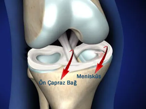
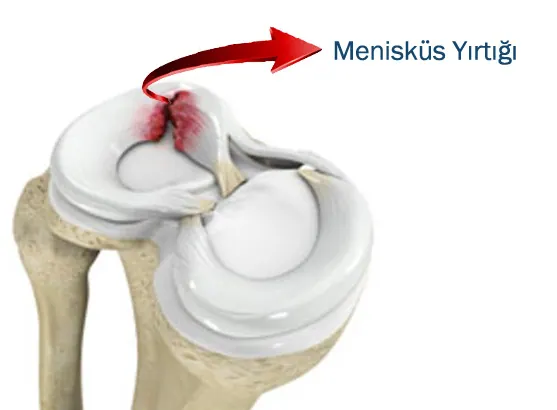
How does a meniscus tear occur?
The knee joint is one of the most frequently injured areas of the human body, and meniscus tears are the most common reason for knee surgery. Contrary to popular belief, meniscus problems do not only affect athletes; they can occur in all parts of society and at any age.
In young people, meniscus tears develop after serious strain such as sports injuries or traffic accidents. When the body twists on a planted foot, the menisci are trapped between the knee bones and tear.
In older people, menisci that are already worn can tear with small movements.
In childhood, congenital abnormalities of the meniscus (such as a discoid meniscus) can lead to early tears.
What are the signs of a meniscus tear?
In young people whose meniscus tears under strain, the patient feels pain, a stabbing or tearing sensation in the knee. Bleeding inside the knee may occur because of the tear, and in that case the knee swells. Within about a month all complaints ease and only point tenderness remains over the tear. If the torn meniscus gets trapped between the bones, the knee locks. In that case the patient cannot bend or straighten the knee. If the knee is forced to move, it may sometimes unlock with a jumping sensation. In both situations the tear is large and requires urgent surgery.
Tears in older patients cause milder complaints. If there is no cartilage wear, the pain is not unbearable. There may be an occasional catching sensation, but locking is rare. Swelling may be seen due to increased fluid inside the joint.
In children with a discoid meniscus, a snapping sensation is felt in the knee. When a tear develops, the pain increases and catching and locking may occur.
How is a meniscus tear diagnosed?
A meniscus tear is diagnosed from how the injury happened, the patient's complaints and the knee examination. With an MRI scan, the location, shape and size of the tear can be determined with 95% accuracy. Injuries to the cartilage, the anterior cruciate ligament and the collateral ligaments that accompany the meniscus tear can also be detected on the knee MRI. Meniscus tears that cannot be detected despite all investigations can be diagnosed by arthroscopic examination.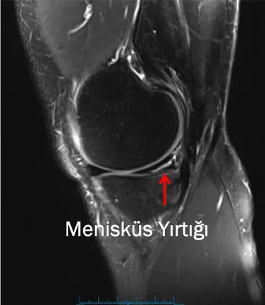
How is a meniscus tear treated?
When there is knee pain after an injury, ice and a bandage are applied early, painkillers are taken and the knee is rested. After that, you should see an orthopaedic surgeon as soon as possible without delay.
Because a torn meniscus usually does not heal on its own, the ideal treatment, especially in young patients, is early surgery. If a meniscus tear is not treated early, serious damage to the joint cartilage can develop over time. As a result, pain and restricted movement may appear in the early period, and in the long term osteoarthritis may develop at a young age due to permanent cartilage damage.
In older patients, surgery may be needed if the knee catches or locks. If these signs are absent, conservative (non-surgical) treatments are used.
Today, meniscus tears are treated with a closed method (arthroscopic surgery).
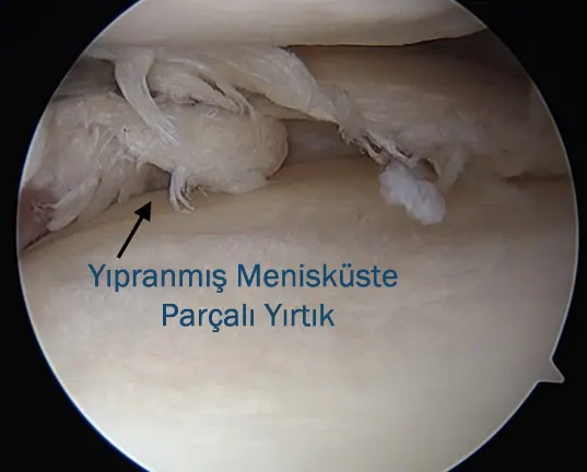
Knee arthroscopy is performed through two incisions of about 0.5 cm. Through one of the two entry holes a thin, tube-shaped fibre-optic instrument called an arthroscope is inserted into the joint, and through the other the instruments used for the surgery. The arthroscope is connected to a screen via a camera. Saline is also given into the joint, both to wash the knee and to keep the image clear. In this way the surgeon sees the inside of the knee clearly on the screen and treats the structures inside the knee.
Meniscus operations that can be performed arthroscopically fall into three groups
1- Arthroscopic partial meniscectomy:
This is the removal of the torn part of the meniscus that causes the problem. It is used for tears in the inner part of the meniscus, where the blood supply is poor, or for worn, fragmented tears that cannot be repaired. Patients can walk on the same day after surgery and return to work within a few days.
2- Arthroscopic meniscus repair:
Because the blood vessels coming from the joint capsule supply the outer third of the meniscus well, tears in this area are suitable for repair. After surgery, depending on the type of tear, the strength of the repair and their occupation, patients can return to work and sport after 6 weeks to 6 months.
3- Meniscus transplantation:
These are methods used in young patients whose meniscus had to be removed. Synthetic menisci (meniscus scaffolds) or menisci taken from a cadaver are used for transplantation. Meniscus scaffolds are used only for partial meniscus loss and are sutured to the removed part of the meniscus. Cadaveric meniscus transplantation is performed in limited numbers in our country and is done when the entire meniscus has been removed. Success at 10-year follow-up is between 60% and 80%.
The information on this page is for general information only and does not replace an examination and diagnosis.